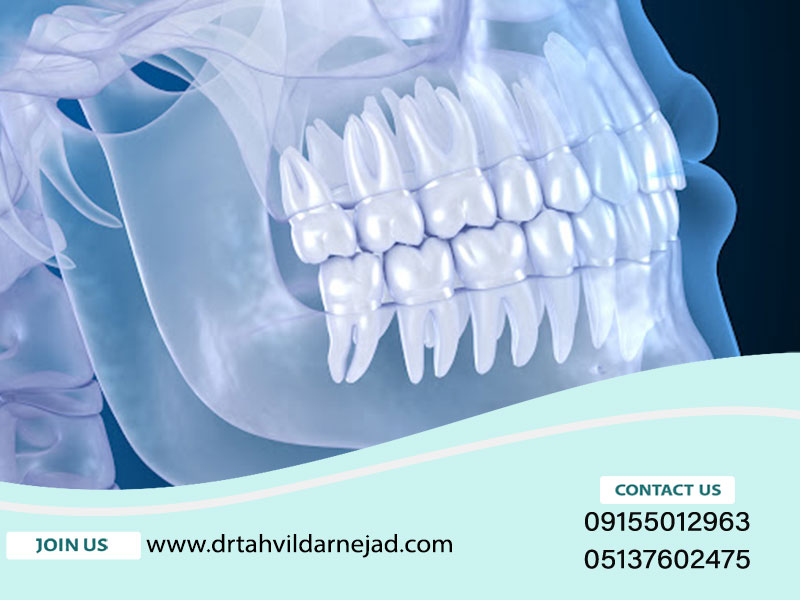
When should we take a dental OPG photo? |Dr. Tardishdarnjad
pspig. ug
OPG (Orthopantomogram) flow アñESe. . �ion�çü‑�� ülliii barrids çççq � � � � � � � � � �� ] � ア �ion�ü‑′� � ���� �������� . p�� � � � � � 0‑DES‒‒‑ . To photograph the teeth with this method, X-type PG photography rays are used, which are emitted by the radiography machine.
But what is the use of this type of photo and how does it help the dentist, we will discuss it in the rest of this article from the official website of Dr.
How does the opg image help the dentist?
In dentistry, as in other medical services, it is necessary to examine the patient's position, damaged tissue, position of teeth, etc. by a dental specialist. so that he can have a proper plan for the treatment and treatment process. In fact, with the help of the OPG photo, the dentist identifies things that cannot be seen in the oral examination. Among the most important of them are:

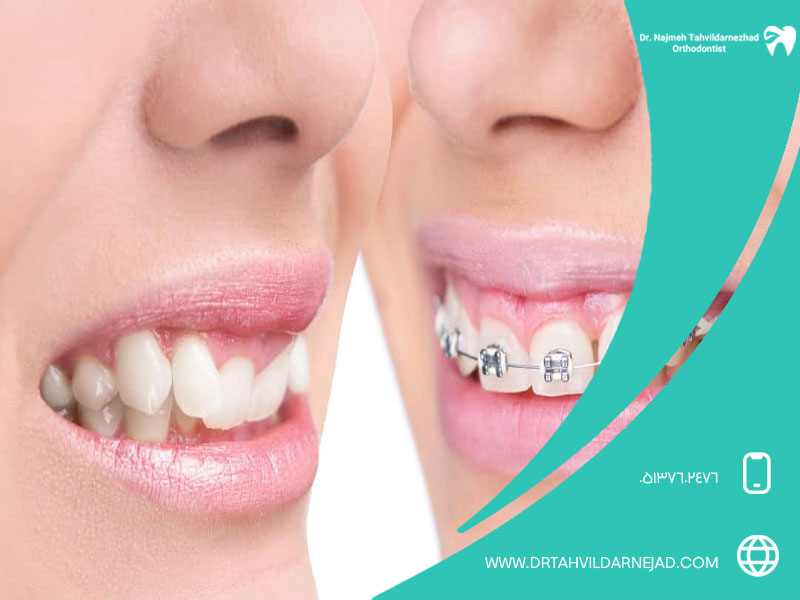
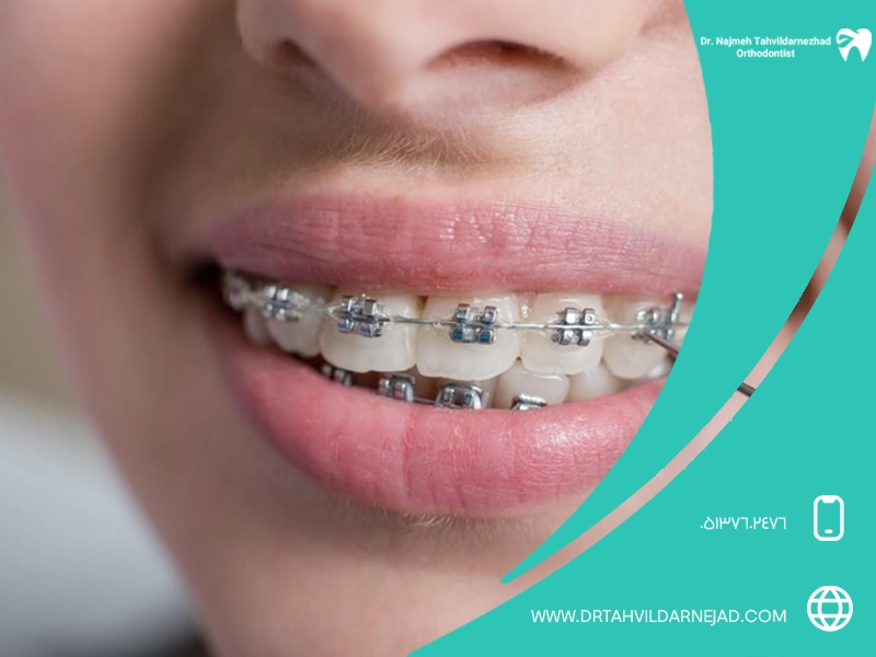
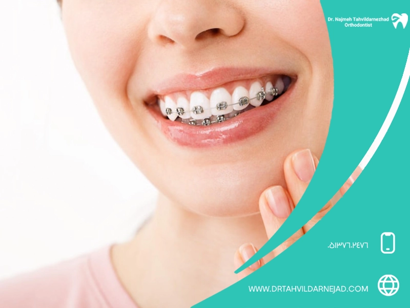
Checking the position of the teeth before orthodontics
The best orthodontic specialist in Mashhad prescribed this photo to fully observe the exact position of the teeth, roots and jawbone. By examining this photo, the orthodontist can set up a detailed and personalized treatment plan for the patient, and among the things that can be seen in this photo, we can refer to the position of the reasons for tooth extraction for orthodontics, hidden teeth, the amount of bone density, the angle of growth of the jaws, and the size of the teeth. OPG is a road map for the orthodontist to choose the best treatment method to correct dental abnormalities.
Diagnosis of cysts in the jaw bone
One of the important uses of OPG is to detect the location of bone cysts. Using this photo, the dentist can determine the exact size, shape and location of the cyst and choose the best treatment method for the patient.
Study of tumors of the jaw and mouth
Another use of OPG photos is to diagnose tumors of the jaw and mouth. By viewing this photo, the dentist can correctly diagnose abnormal changes in the jawbone, such as extra teeth, tumors of the jaw and mouth, etc.
Observation of damaged teeth inside the gums

One of the important uses of this photo is to observe the damaged teeth inside the gums. Using OPG, the dentist can clearly see teeth that are broken, decayed or otherwise damaged, even if they are below the gum line. This helps the dentist to consider a suitable treatment plan for these teeth.
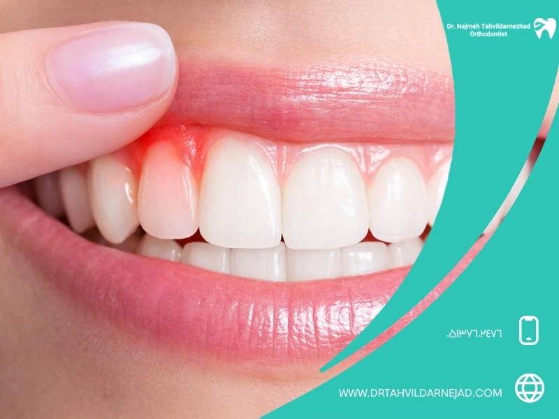
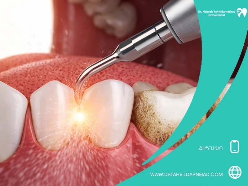
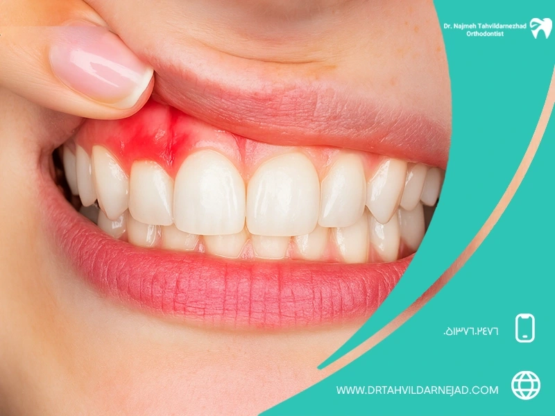
Diagnosis of periodontal disease with OPG photo
OPG photo helps the dentist to evaluate the amount of jaw bone loss caused by periodontal disease. By viewing this photo, the dentist can diagnose the severity of the disease and choose the best treatment method for the patient. In other words, the OPG photo is like an X-ray that allows the dentist to see below the surface of the gums and jawbone, thereby detecting periodontal disease in its early stages.
Read more' Among the things that a dentist recommends OPG photo for a person is to check things like cause of loose teeth. If you want to learn more about this complication, click on the given link.
Examinations before wisdom tooth extraction
Before the dentist performs an operation to extract the wisdom tooth, the position of the tooth, its relationship with the roots of the adjacent teeth, nerves and sinuses are examined. By examining this photo, the dentist can choose the best method for wisdom tooth extraction and avoid possible complications during surgery. In other words, OPG is a road map for the dentist to perform surgery more confidently.
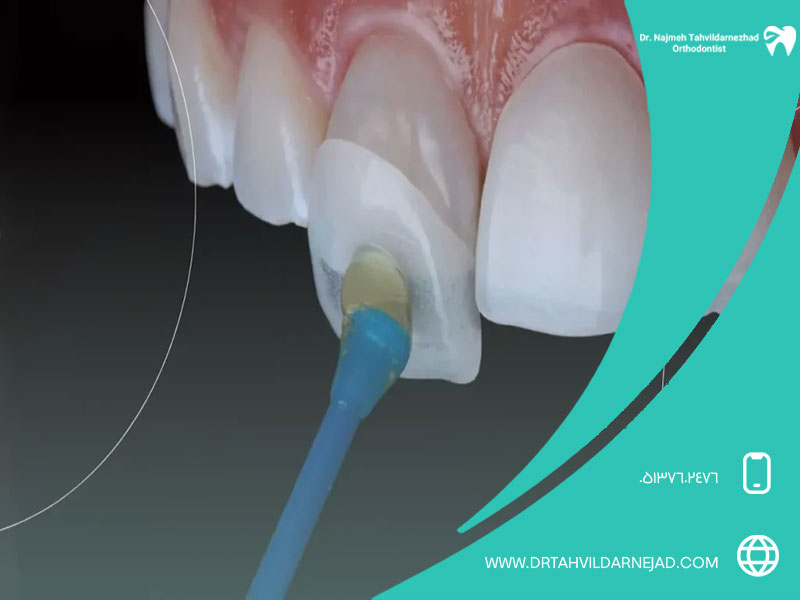
Diagnosis of tooth decay
This photo helps dentists to identify the extent of tooth decay, infections and gum disease. By carefully analyzing the image, dentists can plan appropriate treatments.
Examinations before dental implant implantation

OPG (Orthopantomogram) photo is necessary to check the dental and bone status of patients before implanting dental implants. By using OPG, the dentist can make a more accurate diagnosis and have a better plan for implant placement. After seeing this photo, if a person is facing osteoporosis, he will know can implants be placed with osteoporosis or not.
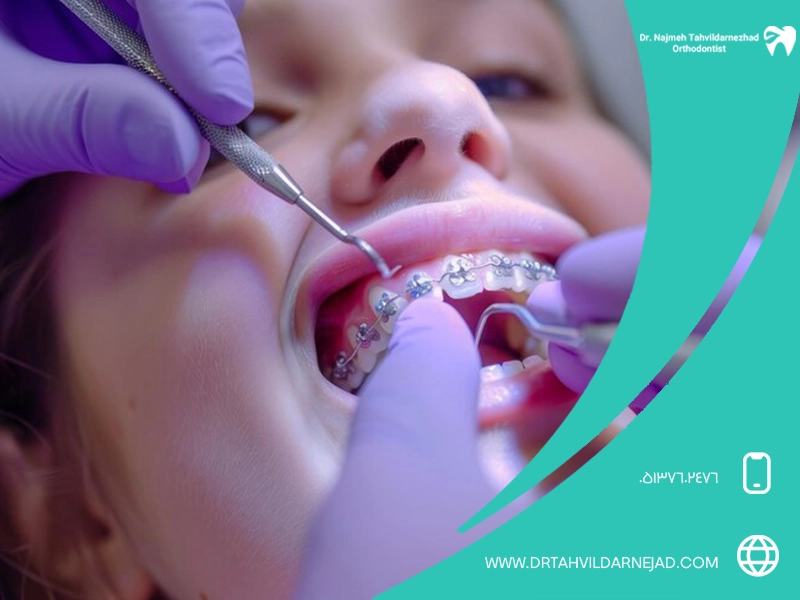
What conditions should we have to take an opg photo of a tooth?
To take dental OPG photos, special conditions are required to perform high-quality and accurate imaging. These conditions include the following:
- Preparation before the photo:
- Before taking the OPG photo, it is better not to wear any metal ornaments on the head and neck such as necklaces, earrings and glasses. Because these objects may disturb the image.
- If you are pregnant or there is a possibility of pregnancy, be sure to inform the dentist.
- If you are allergic to iodine or contrast agents Tell your dentist.
- How to take photos:
- When taking an OPG photo, you must remain still so that the image does not blur.
- You may be asked to open your mouth or put certain objects in your mouth.
- The whole process of taking an OPG photo usually takes less than a minute.
Checking the advantages and disadvantages of OPG
photosOPG or dental panoramic photo provides a general view of the upper jaw, lower jaw, teeth and jaw joints. Its advantages include high speed, low radiation dose and full coverage of the mouth. However, the lower accuracy compared to 3D images and not showing the exact details of the roots are its main disadvantages.
Benefits of imaging OPG
OPG imaging allows simultaneous examination of teeth, jawbone, sinuses and temporal joint. This method is very useful for detecting hidden teeth, evaluating the overall condition of the mouth, and planning orthodontic and surgical treatments. High processing speed and patient comfort are other important advantages of OPG photo.
Disadvantages of OPG imaging
Despite its widespread use, OPG photography has some limitations. This method does not show the fine details of small caries, root cracks or fine lesions very well. Also, there is a possibility of image distortion and reduced resolution in some areas. For this reason, in many cases the need for complementary imaging methods is felt.
What is the difference between photo OPG dental and dental radiology photo?
The OPG image provides a panoramic and general image of the entire jaw and teeth, while conventional dental radiographs, such as periapical or bitewing, have a local and more detailed focus. OPG are used for general examination and local radiology for accurate diagnosis of caries, infection and condition of roots.
Best alternatives to dental OPGshots
In cases where higher accuracy is required, methods such as CBCT, RVG and advanced digital scans replace the OPG photo. These methods provide more details of bone structure, tooth roots and hidden lesions and are more useful in implant treatments, surgery and specialized diagnoses.
Dental digital imaging with RVG | High precision with minimal radiation
RVG is a type of digital intraoral radiography that provides high resolution images and lower radiation dose than traditional methods. This method is very suitable for accurate caries diagnosis, root examination and control of endodontic treatments. High speed and immediate image display have made RVG a popular option in dentistry.
CBCT; The most advanced three-dimensional imaging method of teeth and jaw
CBCT image is an advanced 3D method that provides detailed information of the jawbone, the position of the teeth and the surrounding structures. This method is especially used in implant, jaw surgery and diagnosis of complex lesions. Although its radiation dose is higher than OPG, it has a much higher diagnostic accuracy.
All-round digital panoramic scan | The new generation of dental imaging
Surrounding panoramic scan is a new generation of imaging that provides a more accurate image than traditional OPG with higher resolution and advanced digital processing. This method reduces image errors and is an ideal option for a comprehensive evaluation of the condition of the mouth and jaw, especially in modern dental imaging centers.
Final word
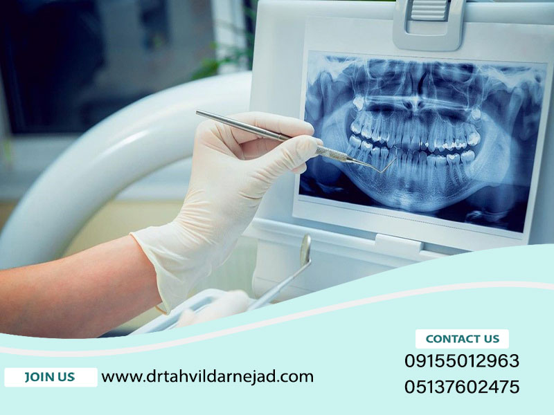
OPG photo (Orthopantomogram) is a type of X-ray photo that records the entire jaws, teeth and surrounding structures in a two-dimensional image. This technique is used to diagnose dental problems, analyze the condition of the jaw, identify hidden teeth, evaluate infections, and analyze bone structures. Also, in planning orthodontic treatments and jaw surgery, OPG is used to check the relationship between teeth and jaws. This picture helps the therapists in making the right decisions and facilitates the treatment process.
FAQ
1- What is an OPG photo?
OPG photo is a panoramic radiograph that shows a general view of the teeth, upper and lower jaw, and jaw joints.
2- When is it necessary to take an OPG photo?
Usually, an OPG photo should be taken to check wisdom teeth, orthodontics, jaw problems, and diagnose gum diseases or cysts.
3- Is it possible to take OPG photos of children's teeth?
Yes, if needed, the dentist also uses OPG to check the growth of permanent teeth or children's jaw problems.
4- What does OPG photo help in orthodontic treatment?
This photo helps the orthodontist to carefully examine the position of the teeth, roots and jaw and choose the best treatment plan.
5- Does the insurance cover the cost of the OPG photo?
Depending on the type of insurance and treatment conditions, some insurances cover part of the cost of this radiography.